---
title: "Part 2: State of Brain Emulation across Organisms — C. elegans"
document_id: sobe-2025-main-ai
document_type: report_section
parent_document: ../report.md
section_id: main-section-06-part-2-state-of-brain-emulation-across-organisms-c-elegans
section_order: 6
section_count: 17
language: en
license: CC-BY-4.0
---
# Part 2: State of Brain Emulation across Organisms — C. elegans
> Selective-retrieval section 6 of 17 from the [complete AI-readable report](../report.md). Content is copied without editorial summarization; relative asset, data, and code paths are rebased for this directory.
- **Parent document:** [`report.md`](../report.md)
- **Section ID:** `main-section-06-part-2-state-of-brain-emulation-across-organisms-c-elegans`
- **Order:** 6 of 17
- **Words:** 7193
- **Previous:** [Part I: Foundations](05-part-i-foundations.md)
- **Next:** [Larval Zebrafish](07-larval-zebrafish.md)
In the following chapters, we juxtapose key anatomical and behavioural features of C. elegans, larval zebrafish, Drosophila, the mouse and the human brain. The former constitute four commonly used animal models in neuroscience. However, many other relevant species exist that this report was not able to cover: from other small fish like Danionella ([Hoffman et al., 2023](https://www.nature.com/articles/s41467-023-43741-x)), bees, ants, cockroaches, rats ([Herculano-Houzel and Lent, 2005](https://www.jneurosci.org/content/25/10/2518); [Welniak-Kaminska et al., 2019](https://journals.plos.org/plosone/article?id=10.1371/journal.pone.0215348), see also these [two](https://youtu.be/9lYPntgZwTI?si=KhxlhjmT9c7PKXNz&t=91) [videos](https://www.youtube.com/watch?v=0jo_EG7XqZQ&ab_channel=NanaBorderCollie)), to marmosets ([Herculano-Houzel et al., 2007](https://www.pnas.org/doi/10.1073/pnas.0611396104); [Seki et al., 2017](https://doi.org/10.1016/j.neuroscience.2017.09.021)) and rhesus macaques ([Dash et al., 2023](https://www.sciencedirect.com/science/article/pii/S0197458023000283); [Herculano-Houzel et al., 2007](https://www.pnas.org/doi/10.1073/pnas.0611396104)) – among others.
The number of neurons varies dramatically across the animal kingdom. Some multicellular animals, like sponges, possess no neurons at all. Among those with nervous systems, counts range from organisms like Intoshia variabili with as few as 4-6 neurons ([Slyusarev et al., 2023](https://onlinelibrary.wiley.com/doi/10.1111/ede.12462)) to elephants with an estimated 257 billion neurons
([Herculano-Houzel et al., 2014](https://doi.org/10.3389/fnana.2014.00046)). Current single-neuron recording technologies allow for monitoring approximately 1,000,000 neurons in mice (~1% of the brain), and a key challenge for the upcoming decade in neuroscience is to scale this to 100 million neurons. However, further increases may eventually encounter physical limitations, potentially capping the maximum number of neurons that can be simultaneously recorded at single-cell resolution ([Marblestone, 2013](https://www.frontiersin.org/journals/computational-neuroscience/articles/10.3389/fncom.2013.00137/full)).
The information presented in the chapters is limited to data and methods that, in theory, allow for single-neuron resolution. Accordingly, barely any non-invasive recording modalities experiments or meso-scale microscopy with resolutions below synaptic resolution are discussed. Comprehensive discussions of general methodological details are discussed in the respective chapters after the organism section. Readers new to the field might want to first read those chapters, as organism chapters assume fluency across Neural Dynamics, Connectomics, and Computational Neuroscience.
Based on outlook sections of papers referenced and conversations with experts, we conclude each organism chapter with a model organism overview and gap analysis:
- Pros and cons of anticipated scientific insights and experimental tractability
- A non-exhaustive list of gaps and illustrative project opportunities
------------------------------------------------------------------------
C. elegans
### Anatomy & Behavior
Caenorhabditis elegans (C. elegans) is a worm the size of a grain of salt (~1mm long, 50 µm in diameter), or 0.002 mm³. Hermaphrodite worms have a total of 300 neurons across their bodies ([White et al, 1986](https://royalsocietypublishing.org/doi/10.1098/rstb.1986.0056), [Skuhersky et al., 2022](https://doi.org/10.1186/s12859-022-04738-3)), with neurons distributed between a dense anterior "brain" region containing roughly half, and the remainder organized in ganglia and as motor neurons throughout the body ([Arnatkevic̆iūtė et al., 2018](https://journals.plos.org/ploscompbiol/article?id=10.1371/journal.pcbi.1005989)). Only a few of these neurons are thought to spike. For example, motor neurons are believed to rely on graded signals rather than firing action potentials, due to their reported lack of voltage-gated sodium channels ([Goodman et al., 1998](https://www.sciencedirect.com/science/article/pii/S0896627300810144)). Specific C. elegans neurons, including sensory types and even certain motor neurons involved in behaviors like defecation, do fire all-or-none, calcium-based action potentials. However, the typical firing rates of these spiking neurons are not yet well characterized across the nervous system, especially under natural conditions. The C. elegans nervous system is known for its overall stereotypy. However, even in this system, studies indicate individual variability in synaptic wiring, with some chemical synapses differing between genetically identical animals ([Witvliet et al., 2021](https://www.nature.com/articles/s41586-021-03778-8)). This variability may contribute to individual behavioral differences. The typical worm lives for roughly two weeks at 20 °C ([Mack et al., 2018](https://www.sciencedirect.com/science/article/pii/S1740675718300343)).
Video 1 - C. elegans behavior [C. elegans responding to a touch stimulus](https://www.youtube.com/watch?v=olrkWpCqVCE&ab_channel=MassachusettsInstituteofTechnology%28MIT%29)
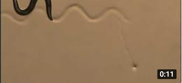
C. elegans exhibits a diverse and adaptive behavioral repertoire, enabling it to thrive in dynamic environments. Its locomotion includes forward crawling, reversals, head movements, and pauses, which combine into higher-order states like "roaming" (rapid movement with few turns) and "dwelling" (slower movement with frequent turns). When food is removed, the worm executes a precise search strategy, starting with local exploration through sharp turns before transitioning to wider-ranging movement. It navigates chemical gradients to locate attractive substances like NaCl and temperature gradients to find its preferred thermal conditions. Remarkably, C. elegans can learn from experience, associating specific cues with food or harm. For example, it increases attraction to odors linked to food and avoids chemicals or temperatures associated with negative experiences. Males display additional behaviors, such as leaving food to search for mates, highlighting the worm’s behavioral flexibility ([Sterling and Laughlin, 2015](https://doi.org/10.7551/mitpress/9780262028707.001.0001); [Bainbridge et al., 2023](https://doi.org/10.1101/688408)).
Beyond wakeful behaviors, C. elegans exhibits sleep-like states, including developmentally timed sleep during larval molting and quiescence in response to stress or starvation. These states involve periods of reduced movement and responsiveness, akin to sleep in other animals. The worm also displays brief spontaneous pauses during feeding and prolonged quiescence after satiation, mirroring behavioral sequences seen in higher organisms. Together, these behaviors – ranging from basic locomotion and navigation to learning, memory, and sleep – underscore the remarkable adaptability of C. elegans and its ability to respond effectively to environmental challenges ([Flavell et al., 2020](https://doi.org/10.1534/genetics.120.303539)).
### Neural Dynamics
#### Neural activity recording
Typical GCaMP-based approach at moderate laser power and short epochs allows repeated short sessions (5–15 minutes each) over several days, totalling a few hours of imaging across the life of a worm without severely compromising the animal. Proof-of-concept whole-brain or whole-body calcium imaging of C. elegans has been achieved, e.g., using light field microscopy ([Prevedel et al., 2014](https://www.nature.com/articles/nmeth.2964)), a fully automated tracking platform to enable freely-moving imaging ([Li et al., 2021)](https://onlinelibrary.wiley.com/doi/10.1002/cyto.a.24483), improved detection, tracking, and segmentation algorithms ([Wu et al., 2022](https://journals.plos.org/ploscompbiol/article?id=10.1371/journal.pcbi.1010594); [Lanza et al., 2024](https://journals.plos.org/plosone/article?id=10.1371/journal.pone.0300628)). Recent advances in live animal labeling, such as NeuroPAL ([Yemini et al., 2021](https://doi.org/10.1016/j.cell.2020.12.012)), create unique fluorescent labels of each neuron in the worm, allowing for almost unambiguous identification of each of the 300 neurons in freely behaving worms, though activity recording itself requires separate, co-expressed functional indicators (e.g., GCaMP). No large-scale whole-body neuron (i.e., recording from 95% of all neurons, across 95% of the volume) activity datasets of C. elegans are available today ([Sprague et al., 2024](https://doi.org/10.1101/2024.04.28.591397); [Simeon et al., 2024](https://arxiv.org/abs/2411.12091v3)). Atanas et al. performed imaging of approximately 60 freely moving animals for a total of ~1,000 minutes of imaging, measuring the activity of approximately 150 head neurons ([Atanas et al., 2023](https://www.sciencedirect.com/science/article/pii/S0092867423008504)). Individual sessions per animal were 16 minutes, often split into two 8-minute segments, and temporal resolution was 1.7 Hz. They used a nuclear calcium indicator (NLS-GCaMP7f) and a spinning disk confocal for volumetric fluorescence imaging, coupled with brightfield imaging of the worm’s behavior. After processing, this generated about 30 GB.
Figure 7 - Overview of the optical neural recording landscape in C. elegans: Radar plots based on the optical recording literature cited in the report. We plot the following dimensions of brain recordings: spatial resolution, brain volume, temporal resolution, and (estimated) individual and cumulative recording duration. The plots split recordings from fixated (A) and freely moving experiments (B). The outer ring is normalized to the maximum known values. Each ring represents one order of magnitude. The data for this figure is available in the [linked data repository](https://docs.google.com/spreadsheets/d/14rglcvdX8Bl6oSWgNnnlOVM_Oot0dHjfFIRHU2Y-mC0/edit?gid=0#gid=0).
A\) fixated

B\) moving

### Machine-readable figure record: `main-fig-07`
```yaml
id: main-fig-07
document_id: sobe-2025-main-ai
document_path: report.md
anchor: main-fig-07
canonical_url: https://brainemulation.mxschons.com/ai/report.md#main-fig-07
figure: "C. elegans optical recording landscape"
assets:
- "../assets/report/main-fig-07a-publication.png"
- "../assets/report/main-fig-07b-publication.png"
data_status: final_circulation_export_raster_with_contextual_current_sources
data_sources:
- "../data/current-repository/recordings/neurodynamics-papers.tsv"
- "../data/current-repository/recordings/neurodynamics-organisms.tsv"
generator: ../code/run_all_figures.py
generator_function: generate_neuro_rec_radar
publication_export_members:
- "images/image34.png"
- "images/image11.png"
canonical_pdf_pages:
- 36
```
#### Inline contextual data (not an exact publication input): C. elegans studies
```tsv
First author Year Organism Fixated / moving Method Number of neurons Temporal resolution: Hz Duration single session min Duration total repeated sessions min Resolution in µm isotropic Individuals studied Brain volume dimensions SNR Perturbation DOI/Link
Kato 2015 C. elegans fixated Calcium 127 1.0 15.0 120.0 1 12.0 https://doi.org/10.1016/j.cell.2015.09.034
Nichols 2017 C. elegans fixated Calcium 108 1.0 15.0 120.0 1 44.0 https://doi.org/10.1126/science.aam6851
Skora 2018 C. elegans fixated Calcium 129 1.0 15.0 120.0 1 12.0 https://doi.org/10.1016/j.celrep.2017.12.091
Kaplan 2020 C. elegans fixated Calcium 114 1.0 15.0 120.0 1 19.0 https://doi.org/10.1016/j.neuron.2019.10.037
Yemini 2021 C. elegans fixated Calcium 125 1.0 15.0 120.0 1 49.0 https://doi.org/10.1016/j.cell.2020.12.012
Uzel 2022 C. elegans fixated Calcium 138 1.0 15.0 120.0 1 6.0 https://doi.org/10.1016/j.cub.2022.06.039
Dag 2023 C. elegans fixated Calcium 143 1.0 15.0 120.0 1 7.0 https://doi.org/10.1016/j.cell.2023.04.023
Atanas 2023 C. elegans moving Calcium 136 1.0 15.0 120.0 1 42.0 https://doi.org/10.1016/j.cell.2023.07.035
Leifer 2023 C. elegans fixated Calcium 122 1.0 15.0 120.0 1 103.0 https://doi.org/10.1038/s41586-023-06683-4
Lin 2023 C. elegans fixated Calcium 8 1.0 15.0 120.0 1 577.0 https://doi.org/10.1126/sciadv.ade1249
Venkatachalam 2016 C. elegans moving Calcium 187 1.0 15.0 120.0 1 22.0 https://doi.org/10.1073/pnas.1507109113
```
#### Inline contextual data (not an exact publication input): organism normalization maxima
```tsv
Organism Total Neuron Count Maximum Neuron Firing Rate (Hz) Average Liftime in Minutes Isotropic resolution Single Neuron recording in µm ref_id supporting_refs ref_note confidence validated_by
C. Elegans 302 10 21600 1 none none
Zebrafish Larvae 100000 80 14400 1 none none
Drosophila 140000 200 57600 1 none none
Mouse 70000000 200 1576800 1 none none
Human 87000000000 415 39420000 1 none none
```
#### Figure-generation source map (valid Python; not standalone plotting code)
```python
GENERATOR_PATH = "../code/run_all_figures.py"
SOURCE_FUNCTION = 'generate_neuro_rec_radar'
SELECTED_SOURCE_LINES = (
'normalized_alpha = 1 - (1 - base_alpha)**(1/num_recs)',
'normalized_alpha = base_alpha',
'alpha=normalized_alpha,',
)
```
While patch-clamp recordings in C. elegans offer millisecond precision for directly measuring electrical activity in neurons and muscles, this technique is practically limited to recording from only one or a minimal number of cells at a time, making it best suited for detailed studies of specific ion channels and synaptic connections. The main drawbacks are that it requires careful dissection of the worm's protective outer layer and can only record from immobilized animals, making it impossible to study neural activity during behavior ([Goodman et al., 2012](https://doi.org/10.1016/B978-0-12-394620-1.00014-X)). Voltage imaging with GEVIs offers a complementary approach, enabling researchers to simultaneously measure electrical activity across multiple cells in living, behaving worms. The main limitation is that GEVIs produce relatively weak fluorescent signals compared to calcium indicators. However, recent advances using rhodopsin-derived indicators, such as Arch-based GEVIs, have improved their brightness and sensitivity ([Hashemi et al., 2019](https://doi.org/10.1073/pnas.1902443116)). Tokunaga et al. recorded voltage in individual neurons from several hundred individuals for 30-60s at 20-250 Hz ([Tokunaga et al., 2024](https://doi.org/10.1038/s42003-024-06778-2)).
#### Neurotransmitters and Neuromodulators
The C. elegans genome encodes over 300 neuropeptides processed from approximately 160 precursor genes, along with around 150 peptide G protein-coupled receptors (GPCRs) ([Taylor et al., 2021](https://pubmed.ncbi.nlm.nih.gov/34237253/)). Recent large-scale deorphanization efforts have identified 461 peptide-GPCR signaling pairs, providing unprecedented insight into the organization of peptidergic signaling ([Beets et al., 2023](https://doi.org/10.1016/j.celrep.2023.113058)). This work revealed that while some peptide-GPCR interactions are highly specific, others display complex combinatorial patterns - individual neuropeptides can activate multiple receptors, and single receptors can respond to various peptides ([Beets et al., 2023](https://pubmed.ncbi.nlm.nih.gov/37656621/); [Ripoll-Sánchez et al., 2023](https://www.sciencedirect.com/science/article/pii/S0896627323007560)). Functional studies indicate that neuropeptides operate across various timescales in C. elegans, from seconds to hours. While typically considered slow modulators, some neuropeptides like NLP-40 can trigger responses within seconds. Recent whole-brain imaging combined with optogenetics has revealed that many rapid functional connections between neurons depend on dense-core vesicle release, suggesting neuropeptides extensively shape fast neural dynamics ([Randi et al., 2023](https://www.nature.com/articles/s41586-023-06683-4)). The peptidergic network appears organized into distinct functional modules, including specialized "hub" neurons rich in dense-core vesicles and highly connected through peptidergic signaling ([Ripoll-Sánchez et al., 2023](https://www.sciencedirect.com/science/article/pii/S0896627323007560)).
#### Perturbation
Due to its anatomy, C. elegans represents an exemplary model organism for perturbation studies. Since the first expression of optogenetic proteins in C. elegans in 2005 ([Boyden et al., 2005](https://www.nature.com/articles/nn1525)), optogenetic studies have revealed numerous insights into neural circuit function ([Piatkevich and Boyden, 2023](https://doi.org/10.1017/S0033583523000033)): neurons controlling locomotor rhythms ([Fouad et al., 2018](https://doi.org/10.7554/eLife.29913)), interneurons that integrate multiple olfactory inputs to represent valence ([Dobosiewicz et al., 2019](https://doi.org/10.7554/eLife.50566)), single neurons that can both encode chemotactic memory ([Luo et al., 2014](https://doi.org/10.1016/j.neuron.2014.05.010)) and regulate multiple behavioral outputs ([Li et al., 2014](https://doi.org/10.1016/j.cell.2014.09.056)), circuits mediating behavioral state switching in response to oxygen levels ([Laurent et al., 2015](https://doi.org/10.7554/eLife.04241)), and specific interneurons controlling chemotaxis programs ([Kocabas et al., 2012](https://doi.org/10.1038/nature11431)). Additionally, optogenetics has revealed how synaptic energy demand regulates glycolytic protein clustering and identified neurons that contribute oscillatory activity controlling backward locomotion ([Piatkevich and Boyden, 2023](https://doi.org/10.1017/S0033583523000033)). Recent technical advances have dramatically expanded the scale and precision of perturbation studies in the worm. Sharma et al. developed TWISP (Transgenic Worm for Interrogating Signal Propagation), a strain expressing both optogenetic actuators and calcium indicators in all neurons while avoiding optical crosstalk –unwanted interference between light signals used for neural activation and activity measurement ([Sharma et al., 2023](https://academic.oup.com/genetics/article/227/3/iyae077/7670367)). Moreover, Randi et al. systematically measured signal propagation between 23,433 pairs of neurons in the worm's head through direct optogenetic activation combined with whole-brain calcium imaging, revealing that actual signal flow often differs from predictions based on anatomical connectivity due to extrasynaptic signaling ([Randi et al., 2023](https://www.nature.com/articles/s41586-023-06683-4)).
### Connectomics
The relative simplicity and high degree of stereotypy in C. elegans neuroanatomy made it the target for the first complete connectome mapping effort ([Brenner, 1974](https://doi.org/10.1093/genetics/77.1.71), [Brenner, 2002](https://www.nobelprize.org/uploads/2018/06/brenner-lecture.pdf)). In 1986, White and colleagues published this landmark reconstruction, detailing approximately 5000 chemical synapses, 2000 neuromuscular junctions, and 600 gap junctions ([White et al, 1986](https://royalsocietypublishing.org/doi/10.1098/rstb.1986.0056)). However, as discussed in later analyses, this connectome was a composite, painstakingly assembled from electron microscopy sections of multiple, different individuals: specifically, three adult hermaphrodites, one L4 larva, and one adult male. This mosaic approach, necessary due to the technical challenges of the time, combined with potential preparation-induced distortions and significant manual curation, resulted in a generalized model. While groundbreaking, this method inherently introduced variability and did not fully capture the precise neuron positions or individual idiosyncrasies now being addressed by modern atlases ([Skuhersky et al., 2022](https://doi.org/10.1186/s12859-022-04738-3)). The underlying assumption of largely consistent neuronal positions and connectivity across individuals ([Varshney et al., 2011](https://doi.org/10.1371/journal.pcbi.1001066)) was nonetheless crucial for this effort.
This initial mapping was later complemented by the reconstruction of the male-specific nervous system, which contains approximately 80 additional neurons primarily involved in mating behaviors ([Jarrell et al, 2011](https://www.science.org/doi/abs/10.1126/science.1221762)). A comprehensive re-mapping effort by Cook et al. (2019) produced updated connectomes for both sexes, correcting earlier errors and introducing synaptic strength weights based on measuring how many consecutive tissue sections each synapse spanned in the electron microscope images ([Cook et al, 2019](https://www.nature.com/articles/s41586-019-1352-7)). More recent work has expanded into developmental neurobiology, with reconstructions across five different developmental stages ([Witvliet et al, 2021](https://pmc.ncbi.nlm.nih.gov/articles/PMC8756380/)), while continuing refinements driven by recent advances in neuron identification, alignment, processing, and scale have improved both accuracy and completeness ([Skuhersky et al, 2022](https://bmcbioinformatics.biomedcentral.com/articles/10.1186/s12859-022-04738-3); [Brittin et al, 2021](https://www.nature.com/articles/s41586-021-03284-x?fromPaywallRec=false)). To date, C. elegans remains the organism with the most individual connectomes reconstructed. This includes approximately ten datasets detailing its complete brain ([Witvliet et al., 2021](https://www.nature.com/articles/s41586-021-03778-8)) or main neuropil ([Brittin et al., 2020](https://doi.org/10.1101/2020.05.24.112870)), complemented by two whole-animal connectomes ([Cook et al., 2019](https://www.nature.com/articles/s41586-019-1352-7)) and a near-complete somatic nervous system wiring diagram ([Varshney et al., 2011](https://doi.org/10.1371/journal.pcbi.1001066)), all from distinct individual specimens. However, due to the highly stereotyped nature of C. elegans neural connectivity, the marginal scientific value of additional individuals may be limited compared to other organisms with more variable nervous systems.
While electron microscopy provided the foundational C. elegans connectome maps, these structural maps alone do not capture the rich molecular complexity that supports the worm's behaviors. Despite having only 300 neurons, C. elegans relies heavily on sophisticated molecular machinery at each synapse, with the worm’s postsynaptic proteins amounting to about half the number for mammals ([Emes et al., 2008](https://www.nature.com/articles/nn.2135)). Optical approaches like expansion microscopy have emerged as crucial complementary tools to understand this molecular complexity. The first C. elegans-specific expansion microscopy protocol (ExCEL) enabled immunostaining for molecular identification and tissue expansion through innovative cuticle permeabilization techniques ([Yu et al., 2020](https://elifesciences.org/articles/46249#s3)). With standard ExCEL achieving 3.5x expansion and iterative ExCEL reaching up to 20x expansion (25 nm resolution), these methods may resolve individual synaptic connections while preserving the critical molecular information that EM cannot capture. However, achieving effective and reliable expansion microscopy in C. elegans remains challenging, with the worm’s cuticle often acting as an impermeable barrier, severely limiting chemical labeling and detection to organs directly exposed to the external environment ([Kuo et al., 2024](https://pmc.ncbi.nlm.nih.gov/articles/PMC11868961/)).
Given a total body volume of 2-6 million μm³ for an adult hermaphrodite worm, imaging at 10nm isotropic resolution would theoretically generate a total of 2-6 × 10¹² voxels. At 1 byte per voxel for a single channel, this would theoretically require 2-6 terabytes of storage, scaling linearly with the number of colors. Historically, early attempts at computer-assisted reconstruction in the 1970s by White et al. highlight the immense computational hurdles of the era. The 'Modular I' computer they employed, a machine with only 64 KB of memory and 22 MB of storage that required custom-written operating systems in assembly language, proved insufficient for the task ([Emmons, 2015](http://dx.doi.org/10.1098/rstb.2014.0309)). Consequently, they resorted to the painstaking manual annotation of printed electron micrographs using colored Rapidograph pens to trace neurons through serial sections. The tracing and reconstruction of the C. elegans required years of painstaking work, with the project taking 15 years to complete. More recent efforts, such as Cook et al., have leveraged specialized software tools like Elegance ([Xu et al., 2013](https://doi.org/10.1371/journal.pone.0054050)), resulting in speedups of several orders of magnitude, and recent advances in machine learning and computational processing have sped up the process further.
### Computational Modelling
The history of C. elegans emulation is marked by ambitious efforts that, while pioneering, were ultimately constrained by the nascent state of neurotechnology and a critical lack of vertical integration. Early conceptual work, such as biophysical locomotion modeling ([Neibur and Erdös, 1991](https://doi.org/10.1016/S0006-3495(91)82149-X)) and planned, though unrealized, comprehensive models in the late 1990s, signaled the field's aspirations. The Virtual C. elegans project ([Suzuki et al., 2005](https://www.fujipress.jp/jrm/rb/robot001700030318/)) made headway by simulating motor control but had to approximate missing biophysical parameters using machine learning, highlighting the data gaps. Later, more comprehensive initiatives like the community-driven OpenWorm ([Szigeti et al., 2014](https://doi.org/10.3389/fncom.2014.00137)), initiated around 2011, and academically-led projects such as [Nemaload](http://nemaload.davidad.org/) (2011-2013), which aimed to leverage the then-new optogenetic tools, also faced these fundamental limitations. At the time, even with the static connectome ([White et al., 1986](https://pubmed.ncbi.nlm.nih.gov/22462104/)) available, the technologies for detailed, dynamic data acquisition were immature. Optogenetic perturbation for causal inference only emerged in C. elegans in 2005. As discussed above, large-scale neural recordings in the worm were, and to a large degree still are, in their infancy ([Stiefel and Brooks, 2019](https://doi.org/10.1007/s13752-019-00319-5)) – partly due to the dynamic nature of the worm's nervous system, which, with its constant motion and deformation, made tracking of individual neurons a challenge ([Nguyen et al., 2017](https://journals.plos.org/ploscompbiol/article?id=10.1371/journal.pcbi.1005517)). This confluence of technological limitations in neural recording, perturbation, tracking, and imaging meant that the kind of comprehensive, correlated dynamic data needed to truly understand and emulate the worm's nervous system simply was not available during those times, directly contributing to the development of Focused Research Organizations (FROs, [Marblestone et al., 2022](https://www.nature.com/articles/d41586-022-00018-5)), designed to be capable of tackling projects requiring such a high degree of systems integration.
Only now, with the advent of high-throughput connectomics, advanced imaging techniques, and increasingly sophisticated computational tools, the field is beginning to amass the data necessary to make meaningful progress towards C. elegans emulation. While no FRO dedicated to C. elegans exists, recent integrative models like BAAIWorm demonstrate important initial successes, such as replicating basic sensory integration and realistic zigzag locomotion behavior ([Zhao et al., 2024](https://www.nature.com/articles/s43588-024-00738-w)). In the following, we discuss the most prominent computational modelling attempts.
#### OpenWorm
OpenWorm, launched in 2011, represents one of the most comprehensive attempts to create an integrative biological simulation of C. elegans, implementing a multi-scale biophysical approach (see [Virtual Worm Project](http://caltech.wormbase.org/virtualworm/) “worm body”) that spans from ion channel dynamics to whole-organism behavior ([Sarma et al., 2018](https://pubmed.ncbi.nlm.nih.gov/30201845/)).
The simulation architecture integrates multiple types of experimental data, though significant gaps remain. Connectome data from electron microscopy provides the basic network structure ([White et al., 1986](https://pubmed.ncbi.nlm.nih.gov/22462104/)), while calcium imaging data informs neural dynamics. However, a major challenge is the limited availability of electrophysiological data - patch-clamp recordings exist for only a small subset of ion channels, necessitating homology-based inference from other organisms for parameter estimation ([Sarma et al., 2018](https://pubmed.ncbi.nlm.nih.gov/30201845/)).
The project's core simulation stack consists of several interconnected frameworks (see figure below), enabling simulation at multiple levels of abstraction - from basic integrate-and-fire neurons to detailed multi-compartment models incorporating Hodgkin-Huxley dynamics ([Gleeson et al., 2018](https://royalsocietypublishing.org/doi/10.1098/rstb.2017.0379)). A critical limitation in the current implementation is the unidirectional flow of information from neural simulation to body mechanics, though work is ongoing to incorporate sensory feedback ([Sarma et al., 2018](https://pubmed.ncbi.nlm.nih.gov/30201845/)).
Figure 8 - Replication from Figure 1 from ([Sarma et al, 2018](https://pubmed.ncbi.nlm.nih.gov/30201845/)). Overview of OpenWorm Simulation stack a) A component diagram describing the relationships between inputs and outputs of sub-projects within OpenWorm b) A highly simplified schematic view of the system of equations executed in the combined c302/Sibernetic system.

### Machine-readable figure record: `main-fig-08`
```yaml
id: main-fig-08
document_id: sobe-2025-main-ai
document_path: report.md
anchor: main-fig-08
canonical_url: https://brainemulation.mxschons.com/ai/report.md#main-fig-08
figure: "OpenWorm stack replication"
assets:
- "../assets/report/main-fig-08.png"
data_status: unavailable_in_repository
publication_export_members:
- "images/image7.png"
canonical_pdf_pages:
- 41
note: "External reproduction after Sarma et al. (2018)."
```
No local numeric plot input was found. The figure is a conceptual diagram, video still, or reproduction from an external publication; its caption and source remain the machine-readable provenance.

The project employs sophisticated validation frameworks, particularly SciUnit, to systematically compare model outputs against experimental data across multiple scales ([Omar et al., 2014](https://raw.githubusercontent.com/cyrus-/papers/master/sciunit-icse14/sciunit-icse14.pdf)). Behavioral validation compares simulated movement patterns against real worm tracking data, while neural validation involves comparing activity patterns with calcium imaging recordings ([Javer et al., 2018](https://www.nature.com/articles/s41592-018-0112-1)). The current implementation can demonstrate basic locomotion, though matching the full repertoire of C. elegans behaviors (including chemotaxis, thermotaxis, and learning) remains a future goal. These limitations reflect computational challenges and gaps in biological understanding of how sensory information is processed to generate behavior.
Figure 9 - OpenWorm simulation: Demonstration of a computational model of the C. elegans. ([Github page](https://github.com/openworm/OpenWorm), 2024)
### Machine-readable figure record: `main-fig-09`
```yaml
id: main-fig-09
document_id: sobe-2025-main-ai
document_path: report.md
anchor: main-fig-09
canonical_url: https://brainemulation.mxschons.com/ai/report.md#main-fig-09
figure: "OpenWorm simulation"
assets:
- "../images/openworm-simulation-animation.webp"
data_status: unavailable_in_repository
publication_export_members:
- "images/image20.gif"
canonical_pdf_pages:
- 42
```
No local numeric plot input was found. The figure is a conceptual diagram, video still, or reproduction from an external publication; its caption and source remain the machine-readable provenance.
The OpenWorm project makes its tools and simulations accessible through the Geppetto visualization platform ([Cantarelli et al., 2018](https://royalsocietypublishing.org/doi/abs/10.1098/rstb.2017.0380)), enabling web-based exploration of models and data. This infrastructure, combined with the project's open-science approach, provides a foundation for community-driven refinement of whole-organism simulations ([Larson, 2021](https://www.youtube.com/watch?v=HlM-vOVEIE8&t=751s&ab_channel=CarboncopiesFoundation)).
#### Simeon et al, 2024
Recent approaches leveraging artificial neural networks (ANNs) offer a complementary data-driven strategy for modeling C. elegans neural dynamics, with particular emphasis on capturing the inherent predictability of neural activity patterns without requiring explicit biophysical modeling ([Simeon et al. 2024](https://doi.org/10.1109/SoutheastCon52093.2024.10500049)). This represents a shift from traditional bottom-up approaches toward letting the underlying structure emerge from experimental data.
Simeon et al.’s approach makes extensive use of calcium imaging datasets, integrating recordings from multiple experimental sources spanning different behavioral contexts - from freely moving to immobilized worms, and from sleep to optogenetically stimulated states. The combined dataset represents neural activity from 284 worms. A key innovation is the standardization of data processing across sources, including z-scoring of calcium signals and temporal alignment, enabling the pooling of data despite varying experimental conditions.
The approach currently focuses on neural activity prediction without direct connection to behavioral outputs. The simulation framework compared multiple neural network architectures, including Long-Short Term Memory (LSTM) networks, Transformer networks, and Feedforward networks. All architectures shared a common structural framework consisting of an embedding layer for neural state representation, a core processing module, and a readout layer for prediction.
Model validation focuses on next-time-step prediction accuracy, using a teacher-student framework where the biological nervous system serves as the teacher and ANN models as students. The approach employs a 50:50 temporal split for validation, where models are trained on the first half of neural recordings and tested on the second half.
Current implementations demonstrate success in short-term prediction of neural activity patterns for up to approximately 20 seconds (accurately predicting roughly 20-30 timesteps of ~0.7s each), with recurrent models (e.g., LSTM) showing superior performance compared to other architectures with the current dataset. However, long-horizon predictive capabilities remain limited. It is worth noting that this represents a prediction of autonomous neural activity without direct modeling of sensory input or behavioral context. The models show consistent scaling properties across different experimental conditions, suggesting they capture fundamental aspects of neural dynamics. CTRNN models, in particular, exhibited the best scaling properties; for instance, their prediction error consistently decreased with more training data, such that doubling the amount of training data reduced the prediction error by a factor of about 1.57. This scaling behavior allows for an estimation of data requirements for longer predictions. To extend predictive success from a baseline of approximately 20 timesteps to the full 180 timesteps the dataset would need to be approximately 30 times larger than the current one. This translates to requiring recordings from roughly 8,400 worms (up from the current 284), highlighting the substantial data requirements for achieving robust long-horizon predictions of autonomous neural dynamics using this approach.
#### Zhao et al, 2024

BAAIWorm, introduced in 2024, represents a significant advance in integrative biological simulation of C. elegans by implementing a closed-loop system bridging brain dynamics, body mechanics, and environmental interactions ([Zhao et al, 2024](https://www.nature.com/articles/s43588-024-00738-w)). The project builds upon OpenWorm's foundational contributions, particularly its cell model morphologies, synaptic dynamics, and 3D body representations, while introducing crucial new capabilities for real-time simulation and behavioral feedback.
The model integrates diverse experimental data, including ion channel dynamics, neural morphologies, electrophysiology, and whole-brain calcium imaging. A notable strength is validating single-neuron models against patch-clamp recordings for five representative neurons (sensory, inter-, command, and motor neurons), with parameters for other neurons derived through functional grouping. The body-environment component leverages detailed anatomical data to construct a biomechanical model comprising 3,341 tetrahedra and 96 muscles.
The neural network model implements 136 neurons as multicompartmental models with sub-2μm compartments, incorporating 14 types of ion channels. Rather than enforcing strict neurotransmitter constraints, the model uses an optimization approach to determine synaptic properties that reproduce observed dynamics. The body-environment simulation employs simplified but efficient hydrodynamics, enabling real-time simulation at 30 frames per second while maintaining behaviorally realistic movement patterns.
The model demonstrates multi-scale validation, from single-neuron current-voltage characteristics to network-level correlation matrices (achieving 0.076 mean squared error against calcium imaging data) and behavioral reproduction of chemotaxis. While the current implementation focuses specifically on zigzag locomotion, synthetic perturbation experiments reveal important insights about neural circuit function, notably, the absence of neurites or synaptic/gap junctions disrupts global neural dynamics and impairs forward motion. A novel Target Body Reference Coordinate System enables precise quantification of movement patterns, providing a stable framework for comparing simulated and biological behavior.
While BAAIWorm represents a significant advance in integrating different scales of biological simulation, several challenges remain, including expanding to the complete 300-neuron network and incorporating additional behaviors beyond chemotaxis.
Figure 10 - BAAIWorm moving towards simulated chemicals. Video showing the simulated worm ([Zhao et al., 2024](https://www.nature.com/articles/s43588-024-00738-w))
### Machine-readable figure record: `main-fig-10`
```yaml
id: main-fig-10
document_id: sobe-2025-main-ai
document_path: report.md
anchor: main-fig-10
canonical_url: https://brainemulation.mxschons.com/ai/report.md#main-fig-10
figure: "BAAIWorm chemotaxis simulation"
assets:
- "../images/baaiworm-chemotaxis-simulation-animation.webp"
data_status: unavailable_in_repository
publication_export_members:
- "images/image3.gif"
canonical_pdf_pages:
- 45
```
No local numeric plot input was found. The figure is a conceptual diagram, video still, or reproduction from an external publication; its caption and source remain the machine-readable provenance.
Figure 11- Replication from Figure 1 from Zhao et al, 2024 BAAIWorm: Data collection, component construction, and model optimization A) Experimental data collection to constrain models. These data include neural morphologies, ion channel models, electrophysiological recordings of single neurons, connectome, connection models and neural network activities. b, The construction of multicompartmental neuron models and connection models (synapses and gap junctions). c, The biophysically detailed C. elegans neural network model without functional neural activities. d, Optimization of the biophysically detailed C. elegans neural network model to achieve realistic network dynamics. Neurons are color-coded to represent membrane potential.

### Machine-readable figure record: `main-fig-11`
```yaml
id: main-fig-11
document_id: sobe-2025-main-ai
document_path: report.md
anchor: main-fig-11
canonical_url: https://brainemulation.mxschons.com/ai/report.md#main-fig-11
figure: "BAAIWorm workflow replication"
assets:
- "../assets/report/main-fig-11.png"
data_status: unavailable_in_repository
publication_export_members:
- "images/image1.png"
canonical_pdf_pages:
- 46
note: "External reproduction after Zhao et al. (2024)."
```
No local numeric plot input was found. The figure is a conceptual diagram, video still, or reproduction from an external publication; its caption and source remain the machine-readable provenance.
### Gap Analysis
In a 2021 Interview, the long-time OpenWorm lead Stephen Larson advocated that the C. elegans emulation ecosystem would benefit massively from an integrative approach, the “Allen Institute for C. elegans research”, that brings the broad range of interdisciplinary experts together ([Larson, 2021](https://www.youtube.com/live/HlM-vOVEIE8?si=cXO9YIN-y-OxXE7w&t=1835)). In 2023, ([Haspel et al., 2023](https://arxiv.org/abs/2308.06578)) proposed systematically determining the input-output functions of every neuron through comprehensive perturbation experiments in order to fully reverse engineer the entire C. elegans nervous system and to simulate its full range of behaviors. The project assumes it will need thousands of hours of experiments, but has not disclosed specific numbers or funding proposals publicly.
A concentrated, interdisciplinary and sustainably funded effort could likely provide conclusive answers to many of the above mentioned points and could mark a transition away from grants aimed at narrow research questions (e.g., genetics, single-neuron knockouts) towards a large-scale integrative project across subject matter experts.
### Table 4 — Model Organism Overview: C. elegans
| Model Organism Overview: C. elegans | Pros | Cons |
|---|---|---|
| Anticipated Scientific Insights | • Completeness Threshold: What anatomical, molecular, and physiological level of detail is sufficient to recapitulate worm behavior in silico? Likely the only current model organism where ultra-high resolution models are possible.
• Allows to determine the relative importance of different variables to overall emulation success: Specifically, non-synaptic signaling in whole-brain models, gap-junctions, deriving function from structure, extrapolation via cell types, etc
• C. elegans promises to be a Rosetta Stone for converting imaging data into functional models. | • Substantial differences to mammalian nervous systems: Some of the worm's neurons primarily use graded potentials rather than action potentials, making direct translation to other organisms challenging (though synapses seem more similar to other organisms). Additionally, it makes particularly heavy use of non- neuronal signaling, which may limit the generalizability.Despite being a model organism in neuroscience and medicine (e.g., longevity research), it is hard to extrapolate from the worm to other organisms.
• Limited behavioral repertoire: While sophisticated for its size, the behavioral repertoire remains relatively constrained compared to vertebrates. Additionally, the worm lacks clear analogues for many higher cognitive functions of interest (though it has many neurotransmitters implicated in human cognitive function).
• Little individualism: The opposite side of a highly conserved organism is that it is hard to study how individual defining characteristics arise. |
| Experimental Tractability | • Substantial existing infrastructure : A decade of OpenWorm and substantial datasets make it the most mature emulation ecosystem of any organism.
• Data acquisition is relatively cheap: The organism is cheaply maintained and can be produced in large numbers. Genetically modifying the organism is possible. No highly specialized equipment is necessary, and C. elegans are relatively cheap to maintain. The scope of all variables is orders of magnitude smaller than in other organisms.
• Simulation is computationally cheap: Memory and compute requirements are easily covered by modern consumer hardware. Everyone can participate.
• Organisms are highly stereotyped: Data can be collected from multiple individuals without getting too many interindividual differences. | • Challenges of Calcium Imaging: The deformable body and brain make tracking for calcium imaging more difficult |
### Table 5 — Gaps and Opportunities: C. elegans
| Gaps and opportunities: C. elegans | Gaps (non-exhaustive selection) | Illustrative Project Opportunities |
|---|---|---|
| Neural dynamics | • Voltage Imaging Technology: Current rhodopsin-based voltage sensors are limited to small fields of view, limiting whole-brain voltage imaging capabilities. Additionally, voltage imaging can detect rapid events like action potentials, but the signal-to-noise ratio remains a challenge, particularly in C. elegans neurons, which often exhibit only small membrane potential changes ( [Hasehmi et al., 2019](https://www.pnas.org/doi/full/10.1073/pnas.1902443116) )
• Calcium Imaging: Nuclear-localized GCaMP, which enables brain-wide recordings, provides more restricted spatial and temporal resolution than other approaches. This affects our ability to capture fast neural dynamics and the precise timing of neural events. ( [Atanas et al. 2023](https://www.sciencedirect.com/science/article/pii/S0092867423008504#sec3) )
• Integration of Multiple Modalities: While some progress has been made in simultaneous calcium and voltage imaging ( [Tokunaga et al., 2024](https://www.nature.com/articles/s42003-024-06778-2) ), current approaches require complex optical setups and careful correction of photobleaching effects. The ability to correlate different measures of neural activity (calcium, voltage, and neurotransmitter release) remains limited.
• Long-term Recording Capabilities: The development of nuclear calcium indicator strains has improved long-term imaging capabilities, but there remains a need for better tools to study neural dynamics across different timescales and behavioral states ( [Atanas et al., 2023](https://www.sciencedirect.com/science/article/pii/S0092867423008504#sec3) ).
• Circuit Manipulation Precision: There is a need for improved tools that allow simultaneous manipulation and recording from multiple identified neurons ( [Piatkevich, 2023](https://www.cambridge.org/core/journals/quarterly-reviews-of-biophysics/article/optogenetic-control-of-neural-activity-the-biophysics-of-microbial-rhodopsins-in-neuroscience/6F9E422992D9418D9418B5F5B5DC5178) ).
• Neuropeptide Signaling Resolution: While recent advances have provided insights into peptide-receptor interactions ( [Beets et al., 2023](https://www.cell.com/cell-reports/fulltext/S2211-1247(23)01069-0?uuid=uuid%3A0cc16903-f1e2-4679-9b88-0d3db5bc2907) ), the technology is still in its infancy, and there is a need for tools to visualize neuropeptide signaling in vivo and understand its temporal dynamics in neural circuit function ( [Watteyne et al., 2024](https://academic.oup.com/genetics/article/228/3/iyae141/7795512#492448382) ). | • Perturbation-free experiments: By automating the handling of individual C. elegans using microfluidics and using a spinning-disk confocal microscopy, labs would perform C. elegans whole-brain imaging of large counts of freely behaving animals.
• Optogenetic perturbation experiments: Activate single neurons, and measure the behavior and changes in all other neurons at using a lightsheet microscope. |
| Connectomics | • Relevance of synaptic variability: Researchers have observed that even for identically aged hermaphrodites, as many as forty to fifty percent of the synaptic connections can differ from worm to worm ( [Brittin et al., 2021](https://www.nature.com/articles/s41586-021-03284-x) ; [Cook et al., 2019](https://www.nature.com/articles/s41586-019-1352-7) ). This means that two worms will share most of the same underlying “scaffolding” (i.e., which neurons physically touch), but their synaptic connectivity at those touches may differ slightly. The relevance of simulation approaches is unclear at this point.
• Gap Junctions : Gap junctions are often incompletely captured. Improved methods are needed for consistent visualization and annotation of electrical synapses across samples. ( [Witvliet et al., 2021](https://pmc.ncbi.nlm.nih.gov/articles/PMC8756380/) )
• Integration of Multiple Modalities: Current approaches typically analyze either connectivity or activity patterns separately. Integrated datasets that combine connectomic reconstruction with activity imaging and behavioral correlates are needed to bridge structure-function relationships.
• Dynamic connectivity : the molecular underpinnings that guide the selective strengthening and formation of specific synaptic contacts, alongside the relative stability of others, remain only partially understood ( [Brittin et al., 2021](https://www.nature.com/articles/s41586-021-03284-x) ; [Cook et al., 2019](https://www.nature.com/articles/s41586-019-1352-7) ). Finally, there is growing recognition that extrasynaptic signaling (including neuropeptides) and gap junctions can be just as critical for circuit function ( [Randi et al., 2023](https://www.nature.com/articles/s41586-023-06683-4) ). However, legacy datasets often have incomplete elements ( [Witvliet et al., 2021](https://pmc.ncbi.nlm.nih.gov/articles/PMC8756380/) ). | • The “definitive” gap junction resolution connectome: A connectome scanned at a resolution sufficient to determine gap junctions reliably.
• The 100 worm connectome project: to study and determine actual variability amongst what are suspected to be highly stereotyped individuals.
• Establish expansion microscopy and protein labelling: ensure this technology reliably works on worms. |
| Computational Neuroscience | • Limited Experimental Electrophysiological Data: Large portions of the worm’s nervous system have only partial biophysical parameterization, i.e., missing parameters like time constants and amplitudes of Hodgkin-Huxley-type conductances of different types, which determine the intrinsic electrophysiological properties of neurons. Likewise, its parameters of synaptic conductances - latencies, rise and decay times, short-term plasticity, EPSP and IPSP (or EPSC and IPSC) amplitudes- are used in simulation approaches. ( [Zhao et al., 2024](https://doi.org/10.7554/elife.31425) ; [Sarma et al., 2018](https://pubmed.ncbi.nlm.nih.gov/30201845/) )
• Limited behavior repertoire and sensory feedback loops: Collect richer behavioral and sensory feedback loop datasets that enable respective modeling and verification of computational C. elegans models.
• Multi-lab Synergy instead of Heterogeneous and uncoordinated datasets: Data sourced for emulation attempts is scattered rather than systematically acquired in standardized formats. This slows the overall process substantially. Although many groups research C. elegans , no lab has all the expertise and technology (e.g., advanced calcium or voltage imaging, computational physics, etc.) required to build a fully integrated simulation. ( [Larson, 2021](https://www.youtube.com/live/HlM-vOVEIE8?si=cXO9YIN-y-OxXE7w&t=1835) )
• Integrate neuropeptide data: Neuropeptides are not accounted for in any simulation approaches.
• Long-Horizon Predictive Capabilities: Current computational brain models are limited to seconds and could be expanded substantially. | • Official C. elegans computational model data Backlog: Concrete list of necessary variables, listed by computational neuroscientists that can be created by students / PhDs / or research groups. This includes electrophysiological parameters as well as behaviors and sensory feedback loops.
• Filling the C. elegans computational model data backlog: Follow-up project to fill in the gaps in electrophysiology and behaviors.
• Proposal for “ C. elegans Emulation Institute”: Integrative proposal / FRO for advancing C. elegans emulation efforts. |