---
title: "Drosophila"
document_id: sobe-2025-main-ai
document_type: report_section
parent_document: ../report.md
section_id: main-section-08-drosophila
section_order: 8
section_count: 17
language: en
license: CC-BY-4.0
---
# Drosophila
> Selective-retrieval section 8 of 17 from the [complete AI-readable report](../report.md). Content is copied without editorial summarization; relative asset, data, and code paths are rebased for this directory.
- **Parent document:** [`report.md`](../report.md)
- **Section ID:** `main-section-08-drosophila`
- **Order:** 8 of 17
- **Words:** 7466
- **Previous:** [Larval Zebrafish](07-larval-zebrafish.md)
- **Next:** [Mouse](09-mouse.md)
### Anatomy & Behavior
The development of Drosophila melanogaster proceeds through four stages: egg, larva, pupa, and adult. The larval stage represents the primary growth period, with a ~200-fold increase in body mass ([Church and Robertson, 1966](https://doi.org/10.1002/jez.1401620309)) and brain expansion from approximately 10,000 neurons ([Jiao et al., 2022](https://elifesciences.org/articles/74968)) to roughly ~130-140,000 neurons in adults. Adult flies at day 7-15 measure 2.5-3 mm in body length and approximately 2 mm in width, with males slightly smaller than females. Under laboratory conditions at 25°C, their mean lifespan typically ranges from 25 to 75 days for females and 25 to 85 days for males. However, this can vary significantly depending on factors like genetics and environment ([Lints et al., 1983](https://doi.org/10.1159/000213136)). The adult nervous system consists of two main parts: the brain and the ventral nerve cord (VNC). The male CNS occupies about 0.054 mm³ ([Berg et al., 2025](https://doi.org/10.1101/2025.10.09.680999)), of which roughly 75% (~0.04 mm³) corresponds to the brain – including the central brain and optic lobes – and about 25% corresponds to the VNC ([Janelia, 2023](https://www.janelia.org/project-team/flyem/manc-connectome)). The adult brain contains ~130-000 ([Dorkenwald et al., 2024](https://www.nature.com/articles/s41586-024-07558-y)) to ~140,000 neurons ([Berg et al., 2025](https://doi.org/10.1101/2025.10.09.680999)), while the male VNC contributes ~33,000 neurons within a ~166,000-neuron CNS ([Berg et al., 2025](https://doi.org/10.1101/2025.10.09.680999)). All neural structures are enclosed by the cuticle – a multilayered exoskeleton partially transparent to visible light and transparent to infrared wavelengths ([Hsu et al., 2018](https://doi.org/10.1101/339531)) – and are oxygenated by the tracheal system, a network of air-filled tubes that branch progressively finer until directly contacting brain tissue.
Neuronal firing rates in Drosophila are diverse. For example, while some sensory neurons fire spontaneously at only 1-2 Hz, others can exceed 200 Hz in response to stimuli ([de Bruyne et al., 2001](https://www.sciencedirect.com/science/article/pii/S0896627301002896), [Dweck et al., 2023](https://doi.org/10.1126/sciadv.adj7032)). However, the brain's overall energy budget limits the average network activity. While comprehensive empirical data on typical, whole-brain average in-vivo firing rates in Drosophila are currently scarce, we can perform a highly approximate, illustrative calculation to explore this metabolic constraint. We come up with a rough estimate of this limit using the measured resting brain oxygen consumption rate of ~160 pmol O2/min ([Neville et al., 2018](https://doi.org/10.1016/j.jneumeth.2017.12.020)), which translates to a total resting ATP budget of ~5.1 x 10¹² ATP/sec (assuming standard energy conversion factors: ~450 kJ/mol O2 oxycaloric equivalent, ~35% metabolic efficiency, ~50 kJ/mol ATP hydrolysis energy). Distributing this budget across ~140,000 neurons using an estimated cost per spike of ~9 × 10⁶ ATP, calculated for a hypothetical action potential in a related blowfly neuron's axon ([Laughlin et al., 1998](https://doi.org/10.1038/236)), yields a maximum average firing rate of approximately 4 Hz. This theoretical upper bound neglects significant synaptic and resting potential energy costs, meaning the actual sustainable average rate is likely lower.
Drosophila melanogaster displays many behaviors ([Kohsaka, 2023](https://doi.org/10.3389/fncir.2023.1175899); [Caldwell et al., 2003](https://doi.org/10.1073/pnas.2535546100)). Larvae show movements like crawling forward and backward, sweeping their head, or rolling over ([Kohsaka, 2023](https://doi.org/10.3389/fncir.2023.1175899); [Clark et al., 2018](https://doi.org/10.1186/s13064-018-0103-z); [Caldwell et al., 2003](https://doi.org/10.1073/pnas.2535546100)). Sensory feedback modulates these movements, allowing larvae to respond to light, physical obstacles, and food availability ([Clark et al., 2018](https://doi.org/10.1186/s13064-018-0103-z); [Busto et al., 1999](https://doi.org/10.1523/JNEUROSCI.19-09-03337.1999)). Adult Drosophila engage in various behaviors, including walking, running, grooming, aggression, mating, and flying. Automated postural analysis has defined over 100 distinct behavioral states ([Berman, 2014](https://doi.org/10.1098/rsif.2014.0672)), with undoubtedly more to be defined in social encounters or the natural environment. Adult flies exhibit a substantially more complex range of movements than C. elegans or larval zebrafish, such as rapid banked turns (body saccades) during flight, which help minimize motion blur and avoid collisions ([Mujires et al., 2015](https://doi.org/10.1242/jeb.114280)).
Video - 3 Fruitfly Behavior
Fighting
A male fruit fly is courting a female fruit fly.
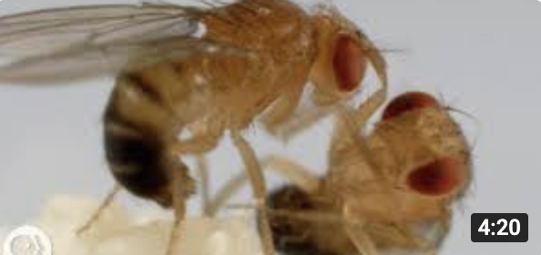
Social mating behavior
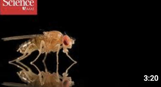
Drosophila demonstrates notable capabilities for learning, memory, and communication. It can habituate to repeatedly presented stimuli (a form of non-associative learning), form aversive courtship memories after rejection, and recall outcomes of past aggressive encounters ([Durrieu et al., 2020](https://doi.org/10.1098/rspb.2020.1234); [Zhao et al., 2018](https://doi.org/10.7554/elife.31425); [Yurkovic et al., 2006](https://doi.org/10.1073/pnas.0608211103), YouTube[ Video](https://www.youtube.com/watch?v=UAF-xo_e_lo&ab_channel=ScienceMagazine)). Social learning also appears in mate choice, foraging decisions, and predator avoidance, with individuals copying behaviors observed in their peers ([Kacsoh et al., 2018](https://doi.org/10.1371/journal.pgen.1007430); [Battesti et al., 2012](https://doi.org/10.1016/j.cub.2011.12.050)). Additionally, even genetically identical flies display stable idiosyncratic “personalities” – for example, consistent left- or right-turning biases and phototactic preferences that persist for days ([de Bivort et al., 2022](https://www.frontiersin.org/journals/behavioral-neuroscience/articles/10.3389/fnbeh.2022.836626/full)).
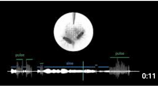
Social behavior is another major facet of Drosophila's ethology and includes aggression, territory defense, and group formation ([Monyak et al., 2021](https://doi.org/10.1242/jeb.238006); [Schretter et al., 2020](https://doi.org/10.7554/eLife.58942)). Both males and females exhibit aggression, although intensity and tactics differ based on sex and experience - males typically engage in higher-intensity boxing and fencing. In contrast, females display more subtle aggressive behaviors ([Zwarts et al., 2012](https://doi.org/10.4161/fly.19249), YouTube[ video](https://www.youtube.com/watch?v=yvd3X1N0jUU&ab_channel=DeepLook)). In some species, territoriality is more pronounced, with males defending leaves or fruit patches, whereas in Drosophila melanogaster, territorial claims tend to be conditional and triggered by factors like female presence or food resources ([Zwarts et al., 2012](https://doi.org/10.4161/fly.19249); [White et al., 2015](https://doi.org/10.1111/evo.12580)). Flies also gather on food sources in aggregations that may improve foraging efficiency and promote social information sharing ([Philippe et al., 2016](https://doi.org/10.1098/rspb.2015.2967)), while maintaining individual spacing that adjusts with density and social context ([McNeil et al., 2015](https://doi.org/10.3791/53242)). Courtship in Drosophila involves an intricate behavior sequence that showcases their social communication capabilities. Males perform elaborate courtship rituals, including following, wing vibration to produce species-specific songs, tapping, and attempted copulation ([Pavlou et al., 2013](https://doi.org/10.1016/j.conb.2012.09.002)). The courtship song, produced by precisely controlled wing vibrations, contains specific patterns of pulses and sine waves that are crucial for species recognition and female choice. These acoustic signals work with chemical cues - males detect female pheromones through specialized receptors, while females evaluate male quality through acoustic and chemical signals ([Fernandez et al., 2013](https://doi.org/10.1007/s00359-013-0851-5); [Lillvis, 2024](https://www.youtube.com/watch?v=fbEMVimwrgQ&ab_channel=HHMI%27sJaneliaResearchCampus)). The successful integration of these multimodal cues - acoustic, chemical, and behavioral - determines mating success and helps maintain species barriers ([Dweck et al., 2015](https://doi.org/10.1073/pnas.1504527112)).
Finally, internal states and social context further influence these behaviors. Isolation can heighten aggression, disrupt sleep and change locomotor activity, suggesting that Drosophila depends on social cues for regulating stress and energy balance ([Eddison., 2021](https://doi.org/10.1038/s41598-021-96871-x); [Li et al., 2021](https://www.nature.com/articles/s41586-021-03837-0)). After mating, females temporarily become non-receptive and increase egg production, a shift mediated by seminal fluid proteins ([Mackay et al., 2005](https://doi.org/10.1073/pnas.0501986102)). Temperature preferences also depend on feeding status – hungry flies often choose cooler temperatures, while sated flies gravitate toward warmth ([Umezaki et al., 2024](https://doi.org/10.7554/eLife.94703.1)). Crowding triggers further behavioral changes, with flies drawing on a sense of "group size" to modify spacing, movement, and interactions ([Rooke et al., 2020](https://www.nature.com/articles/s42003-020-1024-z)). These findings highlight how Drosophila's behavioral repertoire is highly plastic, shaped by physiological, environmental, and social factors.
Neural dynamics
------------------------------------------------------------------------
Figure 13 - Overview of the optical neural recording landscape in Drosophila: Radar plots based on the optical recording literature cited in the report. We plot the following dimensions of brain recordings: spatial resolution, brain volume, temporal resolution, and (estimated) individual and cumulative recording duration. Only recordings from fixated (A) and no freely moving experiments. The outer ring is normalized to the maximum known values. Each ring represents one order of magnitude. The data for this figure is available in the [linked data repository](https://docs.google.com/spreadsheets/d/14rglcvdX8Bl6oSWgNnnlOVM_Oot0dHjfFIRHU2Y-mC0/edit?gid=0#gid=0).
A\) fixated

### Machine-readable figure record: `main-fig-13`
```yaml
id: main-fig-13
document_id: sobe-2025-main-ai
document_path: report.md
anchor: main-fig-13
canonical_url: https://brainemulation.mxschons.com/ai/report.md#main-fig-13
figure: "Drosophila optical recording landscape"
assets:
- "../assets/report/main-fig-13-publication.png"
data_status: final_circulation_export_raster_with_contextual_current_sources
data_sources:
- "../data/current-repository/recordings/neurodynamics-papers.tsv"
- "../data/current-repository/recordings/neurodynamics-organisms.tsv"
generator: ../code/run_all_figures.py
generator_function: generate_neuro_rec_radar
publication_export_members:
- "images/image40.png"
canonical_pdf_pages:
- 65
```
#### Inline contextual data (not an exact publication input): Drosophila studies
```tsv
First author Year Organism Fixated / moving Method Number of neurons Temporal resolution: Hz Duration single session min Duration total repeated sessions min Resolution in µm isotropic Individuals studied Brain volume dimensions SNR Perturbation DOI/Link
Aimon 2019 Drosophila fixated Calcium 63000 100.0 60.0 80.0 1 none https://doi.org/10.1371/journal.pbio.2006732
Schaffer 2023 Drosophila fixated Calcium 40600 10.0 60.0 80.0 1 none https://doi.org/10.1038/s41467-023-41261-2
Brezovec 2024 Drosophila fixated Calcium 95200 1.8 60.0 80.0 1 none https://doi.org/10.1016/j.cub.2023.12.063
Schnell 2017 Drosophila fixated Calcium 10 1.0 60.0 80.0 1 none https://doi.org/10.1016/j.cub.2017.03.004
Aragon 2022 Drosophila fixated Calcium 7500 1.0 720.0 720.0 1 none https://doi.org/10.7554/eLife.69094
Aimon 2019 Drosophila fixated Voltage 7500 200.0 1 none https://doi.org/10.1371/journal.pbio.2006732
```
#### Inline contextual data (not an exact publication input): organism normalization maxima
```tsv
Organism Total Neuron Count Maximum Neuron Firing Rate (Hz) Average Liftime in Minutes Isotropic resolution Single Neuron recording in µm ref_id supporting_refs ref_note confidence validated_by
C. Elegans 302 10 21600 1 none none
Zebrafish Larvae 100000 80 14400 1 none none
Drosophila 140000 200 57600 1 none none
Mouse 70000000 200 1576800 1 none none
Human 87000000000 415 39420000 1 none none
```
#### Figure-generation source map (valid Python; not standalone plotting code)
```python
GENERATOR_PATH = "../code/run_all_figures.py"
SOURCE_FUNCTION = 'generate_neuro_rec_radar'
SELECTED_SOURCE_LINES = (
'normalized_alpha = 1 - (1 - base_alpha)**(1/num_recs)',
'normalized_alpha = base_alpha',
'alpha=normalized_alpha,',
)
```
------------------------------------------------------------------------
#### Neural activity recording
Calcium imaging of small populations of neurons, or all the neurons within a brain region is routine and facilitated by the wealth of genetic tools available in Drosophila. The Drosophila brain's small size and partial transparency suggest the feasibility of whole-brain imaging (i.e., ≥95% of all neurons of ≥95% of brain volume) at single-neuron resolution. In practice, however, the presence of air-filled tracheae creates significant challenges through different mechanisms: for single-photon imaging, light scattering is the primary limitation, while for multiphoton approaches, optical aberrations from the tracheae are the dominant obstacle ([Hsu et al., 2018](https://doi.org/10.1101/339531)). Indeed, while two-photon microscopy can image at depths of 600-800 μm in mouse brain tissue ([Xu et al., 2024](https://www.cell.com/cell/fulltext/S0092-8674(24)00830-4)), it often struggles with imaging depths beyond about 0.1 mm in Drosophila when attempting to image through the intact cuticle or with minimally invasive preparations, largely due to air-filled tracheae. However, with more invasive preparations involving cuticle and superficial trachea removal, functional 2P imaging depths of 120-245 µm ([Aragon et al., 2022](https://doi.org/10.7554/eLife.69094); [Brezovec et al., 2024](https://doi.org/10.1016/j.cub.2023.12.063)) have been demonstrated. Single-neuron resolution in adult Drosophila is also intrinsically challenging because somata are somewhat smaller than in other model organisms. Indeed, typical central brain neurons have soma diameters of 2–6 µm, and mushroom body Kenyon cells are only ~2–3 µm across and densely packed in clusters ([Tuthill, 2009](https://doi.org/10.1523/JNEUROSCI.3348-09.2009); [Gu et al., 2006](https://doi.org/10.1523/JNEUROSCI.4109-05.2006)).
In head-fixed preparations, current techniques navigate these challenges through different trade-offs: light field microscopy achieves fast (~100Hz) imaging of a large portion (600 x 300 x 200 μm3, about ~90% of an assumed 0.04mm³ brain tissue volume) of the brain, though at resolution insufficient to identify individual cells ([Aimon et al., 2019](https://doi.org/10.1371/journal.pbio.2006732)). Schaffer et al. used swept confocally-aligned planar excitation (SCAPE) microscopy to image the dorsal third of the Drosophila brain (450 × 340 × 150 μm³, about ~57% of the assumed brain volume) at single-cell resolution at 8-12 Hz, though scattering and aberrations make resolving individual neurons in deeper brain structures difficult ([Schaffer et al., 2023](https://doi.org/10.1038/s41467-023-41261-2)). This amounted to approximately 1,500 neurons being imaged. Two-photon microscopy offers superior penetration power, but its point-scanning nature creates an inherent trade-off between imaging speed and volume coverage – although advances like light beads microscopy may permit significantly faster imaging ([Demas et al., 2021](https://www.nature.com/articles/s41592-021-01239-8)). Indeed, Gauthey et al. recently demonstrated this potential by achieving whole-brain imaging (e.g., 295 x 675 x 235 μm³, whole brain by bonding box volume) at 28 Hz in preparations where tracheae were surgically removed ([Gauthey et al., 2025](https://doi.org/10.1101/2025.06.18.660371)). This approach, with a spatial resolution of ~1 x 1 x 10 μm, was able to capture fast-timescale auditory responses missed by standard volumetric imaging and could even be pushed to 60 Hz for the central brain alone. Recent work has pushed these limits by imaging the brain (~665 x 333 x 245 μm³, whole brain by box volume) at 1.8 Hz in preparations where the cuticle and superficial trachea were removed ([Brezovec et al., 2024](https://doi.org/10.1016/j.cub.2023.12.063)).
Overcoming the significant optical challenges posed by air-filled tracheae is an important step towards achieving comprehensive whole-brain imaging in Drosophila at single-neuron resolution. Several strategies show promise for mitigating these tracheal effects. Three-photon microscopy is one such avenue due to its inherent advantages in reducing scattering and aberrations ([Hsu et al., 2019](https://doi.org/10.1101/339531)), though it often faces trade-offs with imaging speed. Other approaches, including advancements in light-sheet techniques (like SCAPE or light beads microscopy), the development of improved dissection preparations or optical window techniques that minimize tracheal interference ([Aragon et al., 2022](https://doi.org/10.7554/eLife.69094); [Brezovec et al., 2024](https://doi.org/10.1016/j.cub.2023.12.063)) also offer potential solutions. The goal of these ongoing efforts is to enable consistent, whole-brain functional imaging at single-cell resolution and behaviorally relevant speeds. However, simultaneously achieving optimal depth, speed, resolution, and minimal invasiveness across the entire brain remains a significant technical hurdle. It is worth noting, though, that such advanced imaging may not be strictly required. Indeed, research in Drosophila has typically emphasized specific neural circuits rather than whole-brain imaging, in which defined neural populations express calcium indicators. For imaging of large populations of neurons, researchers generally prefer imaging neuropil despite sacrificing single-cell resolution because it enables consistent registration between specimens. However, structural imaging and reconstruction of the same specimen after functional recording could enable a different approach: imaging the neural cell bodies that form an outer "rind" around the Drosophila brain. This would provide single-cell resolution data while avoiding the need to penetrate deeply through the problematic tracheal system, potentially achieving close to whole-brain functional imaging with 1- or 2-photon approaches ([Harris et al., 2015](https://doi.org/10.1016/j.neuron.2015.05.026)).
Neural recording capabilities are more limited in freely moving or flying Drosophila. Two-photon calcium imaging systems compatible with tethered preparations have been developed ([Seelig et al., 2011](https://doi.org/10.1016/j.bpj.2010.12.735)) and successfully used to record from small numbers of neurons, such as 4-5 pairs of descending neurons ([Schnell et al., 2017](https://doi.org/10.1016/j.cub.2017.03.004)), during wing flapping and steering behaviors that closely mimic natural flight. However, these techniques remain restricted to highly constrained experimental conditions.
Another key challenge is achieving sustained recordings over behaviorally-relevant timescales. Several innovative approaches have recently emerged to tackle this challenge. Huang et al. developed a cranial window preparation that allows repeated imaging sessions over up to 50 days by using laser microsurgery to create and then reseal an optical window in the cuticle ([Huang et al., 2018](https://www.nature.com/articles/s41467-018-02873-1)), while Aragon et al. demonstrated continuous two-photon imaging through the intact cuticle for up to 12 consecutive hours, though this was limited to bright, superficial neurons ([Aragon et al., 2022](https://doi.org/10.7554/eLife.69094)). Most recently, Flores-Valle et al. achieved long-term calcium imaging by collecting 30-second recordings every 5 minutes over 7 days. This intermittent imaging strategy allowed them to accumulate over 15 hours of calcium imaging throughout the week-long experiment ([Flores-Valle et al., 2022](https://doi.org/10.1016/j.jneumeth.2021.109432)). These long-term imaging approaches are, however, typically focused on recording from small numbers of neurons, prioritizing stability and duration over coverage.
Progress is also being made in voltage imaging, where several genetically encoded voltage indicators have been applied to Drosophila brain imaging, including ArcLight, ASAP, Ace2N-mNeon, Varnam, and, more recently, Voltron ([Jin et al., 2012](https://doi.org/10.1016/j.neuron.2012.06.040); [Yang et al., 2016](https://doi.org/10.1016/j.cell.2016.05.031); [Gong et al., 2015](https://doi.org/10.1126/science.aab0810); [Kannan et al., 2018](http://doi.org/10.1038/s41592-018-0188-7); [Abdelfattah et al., 2019](http://doi.org/10.1126/science.aav6416)). Using light field microscopy with the ArcLight indicator, Aimon et al. achieved fast (200 Hz) voltage imaging across a considerable portion of the brain at subcellular resolution, though with fewer extractable activity components compared to calcium imaging due to lower signal-to-noise ratios ([Aimon et al., 2019](https://doi.org/10.1371/journal.pbio.2006732)). Indeed, voltage imaging faces even greater technical challenges than calcium imaging due to poorer signal-to-noise ratios, and the same limitations regarding imaging depth and resolution apply.
#### Neurotransmitters and Neuromodulators
Several GENIs have been successfully validated in Drosophila, including sensors for acetylcholine ([Jing et al., 2018](https://doi.org/10.1038/nbt.4184); [Borden et al., 2020](https://doi.org/10.1101/2020.02.07.939504)), dopamine ([Sun et al., 2018](https://doi.org/10.1016/j.cell.2018.06.042); [Sun et al., 2020](https://doi.org/10.1038/s41592-020-00981-9)), and serotonin ([Wan et al., 2021](https://doi.org/10.1038/s41593-021-00823-7)). A 2009 review identified 119 predicted neuropeptide precursor genes in Drosophila, with 46 neuropeptides biochemically confirmed from just 19 of these precursors ([Clynen et al., 2009](http://doi.org/10.1007/978-1-4419-6902-6_10)); more recent work suggests approximately 50 neuropeptide precursor genes and a similar number of peptide GPCRs ([Nässel & Zandawala, 2019](https://doi.org/10.1016/j.pneurobio.2019.02.003)). In any case, significant progress remains to be made in developing tools to monitor these neuropeptides' dynamics in vivo.
#### Perturbation
The adult fruit fly represents an attractive model organism for perturbation studies, having a well-characterized connectome, a relatively small number of neurons (~140,000), and an extensive genetic toolkit for targeted manipulation. Single-photon optogenetic stimulation using red-shifted opsins like Chrimson is most commonly used in Drosophila, as these wavelengths can penetrate the cuticle without requiring dissection ([Klapoetke et al., 2014](https://doi.org/10.1038/nmeth.2836)). Likewise, individual neurons can easily be optogenetically silenced in behaving flies ([Mohammed et al., 2017](https://pubmed.ncbi.nlm.nih.gov/28114289/)). The powerful genetic toolkit available represents perhaps the strongest aspect of Drosophila neuroscience. The split-Gal4 system enables precise targeting of specific cell types, often achieving single-cell-type resolution that surpasses capabilities in other model organisms ([Meissner et al., 2025](https://elifesciences.org/reviewed-preprints/98405)). This genetic specificity, combined with high-throughput behavioral analysis, allows systematic investigation of neural function across the brain. Upon identification of a neuron of interest, the availability of the connectome and of existing tools permits identification and testing of the role of specific neurons upstream and downstream of that neuron ([Meissner et al., 2025](https://elifesciences.org/articles/98405)). Indeed, these genetic tools have revealed neural populations and activity patterns responsible for a wide range of Drosophila behaviors ([Piatkevich and Boyden, 2023](https://doi.org/10.1017/S0033583523000033)): acquired feeding preferences ([Musso et al., 2019](https://doi.org/10.7554/eLife.45636)), chemotactic navigational decision-making ([Hernandez-Nunez et al., 2015](https://doi.org/10.7554/eLife.06225)), courtship control ([Seeholzer et al., 2018](https://doi.org/10.1038/s41586-018-0322-9)), sleep promotion and locomotor activity suppression ([Guo et al., 2016](https://doi.org/10.1038/nature19097)), long-lasting internal states in the female brain that regulate multiple behaviors ([Deutsch et al., 2020](https://doi.org/10.7554/eLife.59502)), touch signal processing ([Tuthill and Wilson, 2016](https://doi.org/10.1016/j.cell.2016.01.014)), context-appropriate walking programs ([Bidaye et al., 2020](https://doi.org/10.1016/j.neuron.2020.07.032)), complex behavioral sequences ([Vogelstein et al., 2014](https://doi.org/10.1126/science.1250298)), and heading direction representation through ring attractor dynamics ([Kim et al., 2017](https://doi.org/10.1126/science.aal4835)).
Notably, while the recently reconstructed FlyWire connectome provides a complete map of anatomical connections, it does not directly reveal how strongly neurons affect each other's activity in vivo. Pospisil et al. propose that perturbation studies could enable recovery of the "effectome" - a quantitative model of the causal interactions between neurons during brain function ([Pospisil et al., 2024](https://www.nature.com/articles/s41586-024-07982-0)). Their key insight is that while studying all possible pairwise interactions between 140,000 neurons would be intractable, the connectome's extreme sparsity (only 0.01% of neuron pairs form synaptic contacts) provides a powerful prior: neurons without direct anatomical connections are unlikely to have substantial direct causal effects on each other. This dramatically reduces the number of potential interactions that need to be measured through perturbation experiments to recover the fly’s “effectome,” by about four orders of magnitude (∼10⁴-fold) if the approach’s sparsity assumption holds.
### Connectomics
Synaptic resolution electron microscopy efforts in Drosophila achieved an early milestone in 2015 with the complete imaging of a female first instar larva nervous system ([Ohyama et al., 2015](https://www.nature.com/articles/nature14297)), followed by another significant advance in 2018 with the complete imaging of an adult female fly brain using a custom high-throughput serial-section transmission electron microscopy (ssTEM) platform ([Zheng et al, 2018](https://www.cell.com/cell/fulltext/S0092-8674(18)30787-6)). This was followed by focused-ion-beam scanning electron microscopy (FIB-SEM) imaging of the female hemibrain ([Scheffer et al, 2020](https://elifesciences.org/articles/57443)). Subsequent efforts also mapped the female ([Azevedo et al., 2024](https://www.nature.com/articles/s41586-024-07389-x)) and male ([Takemura et al., 2024](https://doi.org/10.7554/eLife.97769.1)) ventral nerve cords from different individuals. Signifying a major step towards holistic maps from single specimens, imaging acquisition has now also been completed for an entire adult male CNS ([Berg et al., 2025](https://doi.org/10.1101/2025.10.09.680999))– a Janelia FlyEM project in collaboration with the Cambridge Drosophila Connectomics Group – and for an entire adult female CNS as part of the BANC project ([FlyWire Blog, 2024](https://blog.flywire.ai/2024/12/20/banc-guide-for-citizen-scientists/)). These initiatives provide the raw data for comprehensive structural maps of individual nervous systems.
Beyond electron microscopy, expansion microscopy (ExM) has emerged as a complementary approach for Drosophila connectomics. When coupled with lattice light-sheet microscopy, ExM has enabled the comparison of neural circuit connectivity across multiple specimens while preserving molecular contrast information ([Gao et al., 2019](https://doi.org/10.1126/science.aau8302)). This protein-specific molecular labeling capability has been further enhanced through transgenic approaches like Bitbow, which enables combinatorial protein barcoding to label neurons uniquely. By targeting five spectrally distinct fluorescent proteins to three subcellular compartments (membrane, nucleus, and Golgi apparatus), Bitbow has demonstrated the ability to generate up to 32,767 distinct molecular barcodes for studying neural circuits in the fly brain ([Li et al., 2021](https://doi.org/10.3389/fncir.2021.732183)). Most recently, advances in expansion microscopy using potassium acrylate-based hydrogels have achieved expansion ratios exceeding 40x, enabling light microscopy visualization of features like mitochondria within presynaptic compartments at resolutions approaching those of electron microscopy while maintaining whole-brain coverage ([Tian et al., 2024](https://doi.org/10.1038/s41467-024-55305-8)). X-ray-based techniques also offer another avenue for large-volume imaging, with X-ray holographic nano-tomography (XNH) achieving 140-170 nm resolution across millimeter-scale volumes of Drosophila nervous tissue ([Kuan et al., 2020](https://doi.org/10.1038/s41593-020-0704-9)).
Assuming a brain volume of 0.04 mm³, imaging at 10 nm isotropic resolution would theoretically generate approximately 4 × 10¹³ voxels. At 1 byte per voxel for a single channel, this would require approximately 40 terabytes of storage per channel.
The progression of Drosophila neuron reconstruction reflects advancing capabilities in automatic neuron tracing and machine-assisted reconstruction proofreading. The initial complete brain imaging by Zheng et al. demonstrated feasibility with a proof-of-concept reconstruction of 120 neurons, with reconstruction requiring, on average, 11.2 person-hours per neuron ([Zheng et al., 2018](https://doi.org/10.1016/j.cell.2018.06.019)). This was followed by a significant improvement in the hemibrain project ([Scheffer et al., 2020](https://doi.org/10.7554/eLife.57443)), which reconstructed over 22,000 neurons and 20 million synapses using automated segmentation supplemented by an estimated 50-100 person-years of human proofreading. The recent FlyWire achievement ([Dorkenwald et al., 2021](https://doi.org/10.1038/s41592-021-01330-0), [Dorkenwald et al., 2024](https://www.nature.com/articles/s41586-024-07558-y)) represents another leap forward, reconstructing the central brain containing 140,000 neurons and over 50 million synapses with 33 person-years of human proofreading, bringing down proofreading time to 19 minutes per neuron ([Dorkenwald et al., 2021](https://pmc.ncbi.nlm.nih.gov/articles/PMC8903166/)). Beyond just the central brain, Azevedo et al. reconstructed approximately 15,000 neurons and 45 million synapses in the ventral nerve cord ([Azevedo et al., 2024](https://www.nature.com/articles/s41586-024-07389-x)), and a fully proofread connectome of an entire adult male CNS has been released ([Berg et al., 2025](https://doi.org/10.1101/2025.10.09.680999)). Further datasets covering the entirety of the fly’s central nervous system are imminent, with the BANC project, also aiming to reconstruct the entire CNS of a single specimen, expected to require several years to complete neuron reconstruction and proofreading ([FlyWire Blog, 2024](https://blog.flywire.ai/2024/12/20/banc-guide-for-citizen-scientists/)).
### Computational Modeling
The relative abundance of connectomics data has profoundly shaped Drosophila brain emulations, from earlier partial reconstructions in FlyCircuit to the recent release of a full adult connectome. This detailed structural knowledge has enabled increasingly comprehensive models, from circuit-specific implementations to whole-brain simulations attempting to capture system-wide dynamics.
#### Huang et al., 2019 and Higuchi et al., 2022
Before a full connectome became available, a few efforts sought to model the whole Drosophila brain by inferring connectivity from the partial FlyCircuit database ([Chiang et al., 2011](https://doi.org/10.1016/j.cub.2010.11.056)). For instance, Huang et al. and Higuchi et al. developed simulations based on ~14-15% of the fly's neurons, employing methods like spatial proximity to estimate connections from morphological data ([Huang et al., 2019](https://doi.org/10.3389/fninf.2018.00099); [Higuchi et al., 2022](https://doi.org/10.1101/2022.11.01.512969)). These groups adopted various modeling approaches: Huang et al. used simplified leaky integrate-and-fire neurons, while Higuchi et al. implemented biophysically detailed Hodgkin-Huxley models, comparing simulated activity to experimental data and known circuit behaviors. Lacking a complete connectome, these early whole-brain models had limited predictive power for the detailed activity of specific neurons or circuits, a challenge subsequent connectome-based models aimed to address.
#### Lappalainen et al., 2024
Another influential example of Drosophila modeling leveraging partial connectomics data came from Lappalainen et al., who developed a "differentiable mechanistic network" (DMN) constrained by anatomical connectivity ([Lappalainen et al., 2024](https://www.nature.com/articles/s41586-024-07939-3)). Their approach combined two electron microscopy datasets spanning different regions of the optic lobe. This connectivity data was complemented by transcriptomics to infer synaptic signs based on neurotransmitter expression, resulting in predictions for both excitatory and inhibitory connections. Since the fly's visual system is organized in repeating columns of similar neural circuits (akin to a convolutional neural network), they used their detailed reconstructions of a small region to build a larger model spanning approximately 45,000 neurons across 721 columns of the central visual field. The team trained their model to detect visual motion using synthetic visual inputs, converting frames from the Sintel film database ([Butler et al., 2012](http://sintel.is.tue.mpg.de/)) into hexagonal arrays of photoreceptor activations that matched the fly's eye structure. Type-specific parameters like membrane time constants, resting potentials, and unitary synapse strengths (a scaling factor for each type-to-type connection, applied to the anatomically measured synapse counts) were optimized for motion detection, specifically, to predict the direction and speed of movement for each point in a visual scene.
Each neuron was treated as a leaky, non-spiking node, with graded synapses encoding excitatory or inhibitory interactions based on known transmitter identities. To translate the network's neural activity into motion predictions, they used a convolutional neural network that took the activity of a subset of neurons as input and produced estimates of movement direction and speed. Model validation involved comparing these motion predictions against ground truth from the Sintel database, alongside testing whether the model reproduced known properties of the fly visual system (e.g., ON/OFF selectivity, T4/T5 direction tuning) established by prior experimental studies. Although training successfully reproduces core visual computations, ablation analyses revealed strong dependence on correct synaptic signs and connectome structure, underscoring the importance of biologically grounded constraints.
#### Shiu et al., 2024
The release of the Flywire reconstruction of the adult Drosophila brain connectome ([Dorkenwald et al., 2024](https://www.nature.com/articles/s41586-024-07558-y)) enabled a new generation of simulations characterized by both whole-brain coverage and connectome-derived structure constraints. Shiu et al. developed one of the first comprehensive models incorporating this data, working with over 127,400 proofread neurons and their 50-plus million synaptic connections ([Shiu et al., 2024](https://www.nature.com/articles/s41586-024-07763-9)). To assign neurotransmitter identities, they leveraged prior large-scale predictions ([Eckstein et al., 2024](https://doi.org/10.1016%2Fj.cell.2024.03.016)), broadly classifying neurons as excitatory (primarily cholinergic) or inhibitory (GABAergic or glutamatergic). Dopaminergic, octopaminergic, and serotonergic neurons were also incorporated and treated as excitatory. Connection weights were derived directly from the Flywire connectome, with sign determined by the assigned neurotransmitter identity. The model required fitting only a single free parameter - the global synaptic weight scale (Wsyn). This global scaling factor parameter determined how strongly each synapse influenced the postsynaptic membrane potential. This single Wsyn value was applied globally, meaning its magnitude was independent of the particular identities of the neurons forming any given connection. It was calibrated using known feeding circuit dynamics, specifically tuned so that 100 Hz activation of sugar-sensing gustatory receptor neurons produced approximately 80% of maximal firing in motor neuron 9 (MN9), a key neuron controlling extension of the fly's proboscis (feeding appendage). This calibration point was chosen based on extensive prior experimental characterization of the sugar sensing in the feeding initiation pathway.
The model employed a leaky integrate-and-fire (LIF) framework with α-synapse dynamics, incorporating synaptic conductance, membrane resistance, and time constants derived from previous Drosophila modeling and electrophysiological studies. Overall, the model made several simplifying assumptions: neurons had zero basal firing rates, gap junctions were not available and thus excluded, and neuromodulatory effects beyond basic excitation/inhibition were not incorporated.
Despite those simplifications, validation efforts yielded encouraging results. Shiu et al. primarily focused on simulating two well-characterized circuits: the feeding circuit and the antennal grooming circuit, to validate their model ([Shiu et al., 2022](https://elifesciences.org/articles/79887), [Hampel et al., 2015](https://elifesciences.org/articles/08758)). They computationally activated subsets of gustatory receptor neurons (sugar, water, bitter, and Ir94e-expressing) for the feeding circuit and analyzed the resulting network activity patterns. Similarly, they simulated the antennal grooming circuit by activating mechanosensory neurons in the Johnston's organ, a sensory structure in the antenna that detects antennal movements. The authors employed three primary approaches to validate these predictions: direct comparison of computationally predicted neural activity with experimental calcium imaging data, in silico silencing experiments to assess the necessity of specific neurons (validated against genetic silencing experiments in real flies), and optogenetic activation studies testing whether specific neurons were sufficient to elicit the behaviors predicted by the model (without embodiment). Through this multifaceted validation strategy, they demonstrated their model could accurately recapitulate known feeding and grooming behaviors, achieving 91% accuracy across 164 experimental predictions and generating novel insights like the inhibitory role of Ir94e neurons in feeding. A companion study further validated the model's predictive power by using it to successfully identify distinct neural circuit mechanisms underlying context-specific halting behaviors in the Drosophila locomotion system ([Sapkal et al., 2024](https://www.nature.com/articles/s41586-024-07854-7)).
#### Cowley et al., 2024
Unlike the connectome-driven approaches described above, Cowley et al. developed their model primarily through behavioral and functional constraints ([Cowley et al., 2024](https://www.nature.com/articles/s41586-024-07451-8)). Their approach centered on understanding how specific neuron types contribute to behavior by systematically silencing neurons and incorporating these perturbations into model training. They collected behavioral data from 459 male-female fly pairs during courtship interactions for model fitting. They recorded six behavioral variables from the male: three movement parameters (forward velocity, lateral velocity, and angular velocity) and three measures of song production (sine song, fast pulse song, and slow pulse song). In each experimental condition, they genetically silenced one of 23 different visual projection neurons (LC) types that form a bottleneck between the optic lobe and the central brain. Their model consisted of three components: a convolutional network processing the male's reconstructed visual experience, a bottleneck layer of 23 units (each representing one LC type), and a decision network producing behavioral outputs. The model processed sequences of 10 frames (~300ms) of visual input to predict behavior, and its training involved "knockout training”: when training on data from flies with a silenced LC type, Cowley et al. set the corresponding model unit's activity to zero, forcing the network to learn how each LC type contributes to behavior. To validate their model, they performed two-photon calcium imaging in head-fixed males from five LC types, testing responses to both artificial and naturalistic visual stimuli. Despite being trained only on behavioral data, their model achieved a 35% correlation with neural responses. The model's prediction that LC types work in combination rather than as independent channels was further supported by analysis of the FlyWire connectome, which revealed shared inputs and outputs among LC types.
#### NeuroMechFly and Vaxenburg et al., 2025
Parallel to these advances in brain simulation, there has also been significant progress in developing detailed embodied simulations of Drosophila. While these models currently employ relatively simple neural controllers compared to the simulations discussed above, they represent an important complementary approach that could eventually enable studying a broader range of fly behaviors when combined with more sophisticated neural models. Two notable efforts in this direction are the NeuroMechFly project and the recent work by Vaxenburg et al. The NeuroMechFly team developed a morphologically accurate model derived from X-ray microtomography data with 65 body segments and 122 degrees of freedom, where each degree of freedom represents an independent type of movement or rotation possible at a joint. The model initially focused on walking and grooming behaviors ([Lobato-Rios et al., 2022](https://doi.org/10.1038/s41592-022-01466-7)). Their first version learned to produce stable walking patterns matching those seen in real flies – see the [video](https://static-content.springer.com/esm/art%3A10.1038%2Fs41592-022-01466-7/MediaObjects/41592_2022_1466_MOESM12_ESM.mp4) – including the typical three-legged walking pattern where legs move in alternating groups of three. Their 2024 update expanded the model's capabilities to include vision and smell, demonstrating more complex behaviors like following scent trails while avoiding obstacles in their path ([Wang-Chen et al., 2024](https://www.nature.com/articles/s41592-024-02497-y)). Notably, this updated version integrated the connectome-constrained model developed by Lappalainen et al. to simulate visual processing during a fly-following task. Vaxenburg et al. took a different approach, building their model from high-resolution confocal microscopy data with 67 body segments and 102 degrees of freedom, along with sophisticated physics modeling including fluid dynamics for flight and special features that allowed the simulated fly to stick to surfaces like real flies do ([Vaxenburg et al., 2025](https://doi.org/10.1038/s41586-025-09029-4)). Through reinforcement and imitation learning, their ANN-driven model successfully replicated both walking and flight behaviors from real fly trajectories, including complex maneuvers like rapid turns and sudden changes in direction. Their model also demonstrated behaviors such as maintaining altitude over uneven terrain and navigating through winding trenches without collision.
### Gap Analysis
Like larval zebrafish, Drosophila represents a unique convergence of experimental tractability and biological sophistication, making it an attractive target for integrated brain simulation efforts. Scheffer and Meinertzhagen recognized the need for integrated approaches in a comprehensive 2021 analysis, where the authors outlined 15 key areas requiring coordinated investigation beyond connectomics, spanning biochemistry, cell physiology, and whole-animal concerns ([Scheffer and Meinertzhagen, 2021](https://doi.org/10.1242/jeb.242740)).
A core technical challenge facing Drosophila-based approaches stems from challenges of imaging all neurons simultaneously. The presence of air-filled tracheae and fat deposits in Drosophila creates significant challenges for whole-brain imaging at cellular resolution. However, the ability to efficiently image defined populations of neurons, as well as easily perturb those defined neurons while measuring both neural activity and behavior, makes Drosophila a powerful model for neuron modeling.
Similar to larval zebrafish, adult Drosophila has only minor clinical and industrial applications. However, fruit flies do represent stable adult-stage organisms rather than rapidly developing larvae. As a result, they could potentially support neural recording in the same individuals over longer time horizons, without the complications caused by rapid neural development. This eliminates many of the constraints that characterize larval zebrafish work, where the brief developmental window limits experiment duration and complicates data integration across modalities. Furthermore, adult Drosophila exhibit a substantially richer behavioral repertoire, including sophisticated social behaviors. This behavioral sophistication could provide more stringent validation criteria for whole-brain Drosophila simulations, including better tests of out-of-domain generalization.
### Table 8 — Model Organism Overview: Drosophila
| Model Organism Overview: Drosophila | Pros | Cons |
|---|---|---|
| Anticipated Scientific Insights | • Rich Adult Behavioral Repertoire: Complex social behaviors, learning, individuality and motor sequences provide sophisticated validation targets.
• Structure-Function Bridge: Could illuminate approaches to inferring causal models in systems where exhaustive recording/perturbation is not feasible. | • Evolutionary Distance: Further from mammalian brain architecture than other tractable models like larval zebrafish. |
| Experimental Tractability | • Mature Research Ecosystem: Drosophila has a broad research community, with sophisticated genetic tools including frontier technologies such as facile single cell-type specific genetic access, combinatorial barcoding libraries already being close to achieving whole-brain coverage (Bitbow's 32,000+ barcodes) and expansion microscopy protocols achieving electron microscopy-like spatial resolution (>40× expansion with Re-PKA-ExM), and was able to leverage this for larger consortia.
• Multiple Technical Paths Forward: Complementary advances in EM, ExM, and X-ray microscopy provide diverse routes to molecular and structural mapping
• Stable Adult Platform: Unlike larval models, it allows extended experiments and complex behavioral studies
• Practical Advantages: Low-cost maintenance and manageable computational scale (~140k neurons)
• Feasibility–Complexity Sweet Spot: ~140k neurons, still small enough for whole-brain connectomics and near-whole-brain imaging. | • Challenges performing whole-brain imaging. Compared to the transparent larval zebrafish brain, high speed, whole brain imaging at cellular resolution is more challenging.
• Behavioral Recording Constraints: Current imaging setups (head-fixation, etc.) somewhat limit the observable behavioral repertoire, particularly for organisms capable of complex behaviors like courtship or flying. |
### Table 9 — Gaps and Opportunities: Drosophila
| Gaps and opportunities: Drosophila | Gaps (non-exhaustive selection) | Illustrative Project Opportunities |
|---|---|---|
| Neural dynamics | • Imaging Trade-offs: Current approaches navigate these challenges through different compromises. Light field microscopy achieves fast (~200Hz) whole-brain imaging but at sub-neuropil resolution. Two-photon microscopy provides cellular resolution but usually requires head fixation and has a limited field of view.
• Advance the frontier of maximum recording time in Drosophila : Currently, up to ~15h are possible. Given that the flies can get up to 75 days old, potentially up to an order of magnitude more is theoretically possible.
• Neuromodulation in Drosophila : bigger datasets on neuromodulation are needed | • Deep-Brain Three-Photon Performance: Systematically characterize resolution, signal-to-noise ratio, and photodamage limits of three-photon microscopy in deepest regions of the Drosophila brain. Establish performance benchmarks for imaging through trachea-filled tissue at cellular resolution.
• Whole-Brain Voltage Dynamics : Optimize next-generation voltage indicators and imaging preps for simultaneous recording from >10,000 neurons across multiple Drosophila brain regions at millisecond resolution.
• Develop some way of imaging flies in complex behaviors , including extremely lightweight threads for continuous brain recording.
• Expand single brain recording horizon: Develop sophisticated repeated imaging abilities for fruit flies that allow for 25 h+ of recording per individual |
| Connectomics | • Molecular Limitations: The FlyWire connectome, derived from electron microscopy, provides primarily morphological and connectivity information, in addition to limited data to distinguish excitatory vs. inhibitory neurons,, lacking crucial details about neurotransmitter identities, receptor distributions, and other molecular properties that influence circuit function. The integration of barcoding and ExM approaches towards creating a comprehensive ExM-derived molecularly annotated connectome is nascent, with some pioneering work at Janelia.
• Inter-individual Variability: While there are indications of stereotypy in the fly brain's neural circuits, systematic understanding remains limited as the community has reconstructed little more than “one and a half" connectomes to date. Moreover, it does not yet account for sex differences. | • Aligned neuronal recordings and connectomics: Ideally, both males and females will reconstruct whole CNS connectomes, combined with extensive calcium imaging prior to reconstruction.
• Whole Body EM: Scan the whole organism and trace nerves with more detail across the organism.
• Multiplexed Molecular Mapping Protocol: Develop a protocol for iterative antibody labeling and imaging compatible with high-expansion (>40×) ExM and enable reliable detection of 15+ proteins through serial rounds of staining.
• Synaptic-Scale X-ray Microscopy: Given that Drosophila has some of the finest neurites known to exist, demonstrating sufficient resolution imaging of Drosophila brain tissue using X-ray ptychography and validating against electron microscopy ground truth could pave the way for future X-ray-based whole-brain imaging applications. |
| Computational Neuroscience | • Missing Molecular Foundation: While the FlyWire connectome provides complete structural connectivity, current models must bridge a significant structure-to-function gap through multiple assumptions, including neurotransmitter identities, synaptic strengths, and more (Eckstein et al., 2024).
• Integrated Neuromechanical Framework: Current tools typically separately simulate neural activity and biomechanics. To integrate these components, unified simulation platforms are needed.
• Limited Experimental Electrophysiological Data: Electrophysiological data (e.g., membrane potentials or spiking frequency) is often the most useful inputs and outputs for computational modeling, yet this data is challenging to collect in Drosophila . | • Resting state Drosophila brain model: There still has not been a full Drosophila brain simulation in which all neurons would be active at least at their spontaneous levels across the whole brain. One needs a spontaneously active brain network within which some sensory or other signals can be instantiated.
• “Emulate the fly” roadmap: An end-to-end plan for collecting all components to create a compelling emulation of the fly.
• OpenFly framework: An effort that aggregates all the computational neuroscience data and provides computational tools for everyone. |